
Now 3D printing which started a few decades ago, has kept this name albeit not a printing act anymore but a construction for which a more appropriate term would be welcome.
In fact, the access to that third dimension is opening new horizons in many fields and will certainly develop in domains that are, still now, unimaginable, such as architecture.
Yes, as it is just a matter of scale and proper building material, we shall see, together with technologies yet unknown, and others already developping such as automation and robotics, the birth of new concepts.
Already, at smaller scales, we see materials being used and adapted to various purposes from the traditional sculpture, to biological matter, while on the other hand, research on stem cells allows us to foresee new domains that, yesterday, belonged to science fiction.
In 1962, Marshall Mc Luhan published his book "The Gutenberg Galaxy" in which he brilliantly forecasts the prevalence of electronic media to the detriment of printed media.
In fact what he calls "the Marconi galaxy" developped widely even though printed matter was still going on and developping as well.
He regarded wireless means of communication (and therefor created the word "media") as the future, a new era.
Little did he know (and could not imagine) the wide spread of computers, personal computers and the tremendous explosion of digital technologies.
Neither could-he conceive the birth of 3D!
Text by © Armand Dauré
More about him:
The Gutenberg Galaxy en.wikipedia.org

De humani corporis fabrica libri septem (On the fabric of the human body in seven books) is a textbook of human anatomy written by Andreas Vesalius (1514–1564) in 1543.
Printable organs
Using modified inkjet printers into 3d printers, scientists are producing three-dimensional living biological tissue. The printer cartridges are washed out and filled with a suspension of living cells and a "smart gel". Alternating patterns of the smart gel and living cells are printed using a standard print nozzle. The cells fuse together to form tissue, and tube formation has been demonstrated with ovarian hamster cells. When finished, the gel is cooled and washed away, leaving behind only the live cells.
The gel is heat sensitive - solidifying at 32 degrees celsius and liquifying at 20 degrees
The gel is heat sensitive - solidifying at 32 degrees celsius and liquifying at 20 degrees
What is the Bio-Printing?
The bio-printer is the product of nScrypt Inc. It is a fully computer-controlled delivery device. Three-dimensional printing is achieved by the movable x-y stage and three z-directional printing heads. Two of these are used to print the bio-ink particles, which are extruded from a bio-cartridge (a micropipette filled with bioink particles) by the positive displacement of a piston within the micropipette. The third unit is pressure operated and is used to print the bio-paper/substrate (e.g. collagen gel). Each extruder is equipped with a camera, providing full visual control of printing.

What is the Tensiometer?

The parallel plate compression device allows determining tissue surface tension and characteristic elastic and viscous parameters. A spherical cell aggregate (A) is placed between the lower (LCP) and upper (UCP) compression plates, in the inner chamber filled with tisssue culture medium. A water jacket, the outer chamber (OC) heats up the system to maintain 37C physiological temperature. The upper plate hangs from a nickel-chromium wire (NCW) attached to an electrobalance (B) that monitors the force applied to the aggregate. Raising the lower compression plate compresses the cell aggregate. This deformation is maintained throughout the compression to measure force dissipation under constant strain. Equilibrium is reestablished through a biphasic relaxation process; the equilibrium force and the geometric parameters are used to calculate the tissue surface tension via the Laplace-Young equation. Viscoelastic properties are determined employing the full relaxation curve.
What is the Magnetic Tweezers?
The magnetic tweezers allows measuring intracellular and cell-level viscoelastic parameters. It is a 2-coil design, capable of generating a constant magnetic gradient (and implicitly a constant magnetic force) over a surface exceeding 4x10⁵ square microns. It is a miniaturized Faraday balance mounted on the stage of an inverted Olympus IX-70 microscope. The force (in the range of 1-1000 pN) is applied unidirectionally, in the horizontal plane, through paramagnetic beads attached to the biological sample. The bead motion under magnetic force is recorded and the trajectory is determined with sub-pixel accuracy by an in-house developed particle tracking program. Physical parameters are determined from the analysis of the bead trajectory.

How to print a human organ?
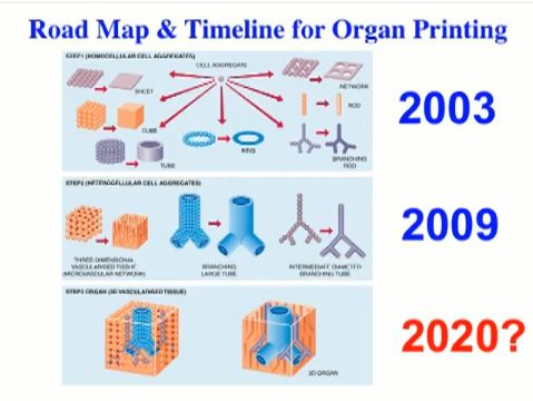
Organ Printing: Future of Rapid Prototyping in Tissue Engineering
How to print a organ by Vladimir Mironov
BBC News
Dr. Vladimir Mironov explains organ printing technology. (2009)
US researchers at Cornell University have engineered an ear made of silicone using a 3D printer, which they hope will one day be capable of producing functional human body parts.
LINKS:
Hod Lipson: mae.cornell.edu
No comments :
Post a Comment
Welcome to 3D TODAY!